
Hypselodoris koumacensis
Anatomy
PHOTO
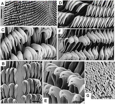
UPPER: Scanning electron micrographs of the holotype of H. koumacensis. A, Section of radula showing central region and left side; B, central region; C, inner laterals on left side; D, mid-laterals on left side; E, inside of outermost left laterals, F, outermost left laterals;
G, jaw rodlets. A, scale = 200 mm; B-G, scale = 20 mm.
Return to H. koumacensis [= H. kaname]
BUCCAL ARMATURE: The radular formula of the holotype, 32mm long preserved, is 98.0.98 x 65. All the teeth, except for the outer few are of similar size. The innermost lateral tooth on each side has a recurved tricuspid blade. The innermost cusp is relatively short and straight, approximately half the length of the other two, which are slightly curved. Of these two cusps, the outer is somewhat longer. Tooth 2 is very similar in shape to Tooth 1 except it lacks the small inner cusp. No outer denticles are present. From Tooth 2-60 the teeth are all of similar shape and size although the cusps become progressively longer. From Tooth 60 the whole tooth begins to gradually diminish in size and the cusps become shorter. From approximately Tooth 80 the outer of the two cusps becomes relatively straight and slightly shorter than the curved inner cusp. At approximately Tooth 90, one small outer denticle appears on the outer edge of the blade, and within two or three teeth there are four or five outer denticles. The outer two or three teeth are reduced to triangular plates. The jaw rodlets are unicuspid.
REPRODUCTIVE SYSTEM: The short muscular vagina leads to a large spherical gametolytic sac. The exogenous sperm sac is reduced to a minute diverticulum off the vagina near its opening into the gametolytic sac. The exogenous sperm duct opens from the vagina opposite the exogenous sperm sac opening. This duct is relatively long and folded and runs to the fertilisation chamber in the female gland mass. The penial sac is slightly longer than the vagina and joins to the large prostate gland by a short muscular vas deferens. The vestibular gland consists of a layer of tubular glands lying along the side and ventral surface of the female gland mass.
This description and figures are from the original description of H. koumacensis:
• Rudman, W.B. (1995) The Chromodorididae (Opisthobranchia: Mollusca) of the Indo-West Pacific: further species from New Caledonia and the Noumea romeri colour group. Molluscan Research, 16: 1-43.
Rudman, W.B., 2001 (May 3) Hypselodoris koumacensis Anatomy. [In] Sea Slug Forum. Australian Museum, Sydney. Available from http://www.seaslugforum.net/factsheet/hypskoumanat
