
Haminoea zelandiae
(Gray, 1843)
Order: CEPHALASPIDEA
Superfamily: HAMINOEOIDEA
Family: Haminoeidae
DISTRIBUTION
New Zealand
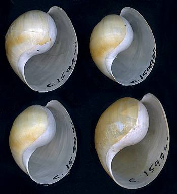
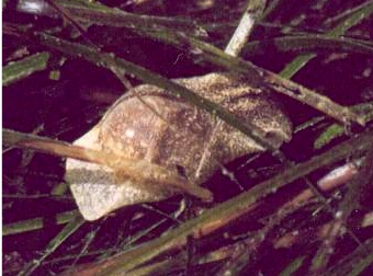
PHOTO
Upper: Herne Bay, Waitemata Harbour, Auckland, New Zealand. Intertidal pools ony rocky reef, 26 September 1968. [Rudman 1971, J. Nat. Hist. 5: 647-75]. Shell length to 22 mm. AM C159944. Photo: Alison Miller & Bill Rudman
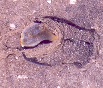
Lower: Black animal crawling through silty sand. Tauranga Harbour, east coast, Nth Island, New Zealand, April 2003. Photo: Paul Furneaux.
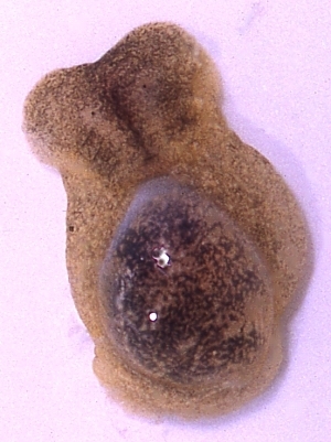
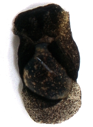
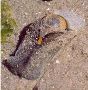
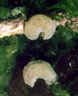
The animal is typical for the genus with a large headshield, slightly tapering and bilobed posteriorly. The parapodia fold up to enclose much of the shell. The foot extends back about halfway along the length of the shell. A lobe of the mantle extends posteriorly from the shell to form a posterior 'pseudofoot' and also a fold which encloses the posterior end ['spire'] of the shell. The animal is translucent with a very variable colouring. Some animals are very pale in colour with mottled specks of brown, black and yellow while others are almost uniformly black or brown. There is a whole range of colour patterns between these two extremes.
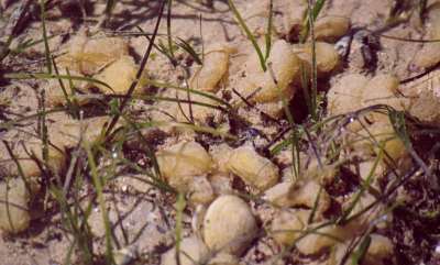
It is found intertidally throughout the North Island and northern part of the South Island of New Zealand in sheltered bays and harbours. It is often found in large numbers on both soft and hard shores, equally at home in sandy mud and in coralline turf on a rock platform. It feeds on green algae such as Ulva and Enteromorpha and also scrapes the diamotaceous film from sea grass and muddy sediments. Its sausage-shaped yellow egg masses can be extremely abundant during the breeding season. It grows to approx 30mm long with a shell length of approx 20mm.
Shell ovate, rather more convex and inflated than most species of the genus. The shell is very thin, fragile, and translucent white. There is often a yellowish periostrical layer. The inner lip often forma a broad callus over the columella
.
See description of gizzard plates below.
Reference:
• Gray, J.E. (1843). Catalogue of the species of mollusca and their shells which have hitherto been recorded as found at New Zealand, with the description of some lately discovered species. In: Dieffenbach, E. Travels in New Zealand with contributions to the Geography, Botany and Natural History of that country. Vol II. Fauna of New Zealand. pp228-263.
• Rudman, W.B. (1971): On the opisthobranch genus Haminoea Turton & Kingston. Pacific Science, 25(4): 545-59, 12 figs.
• Rudman, W.B. (1971) Structure and functioning of the gut in the bullomorpha (Opisthobranchia) Part 1. Herbivores. Journal of Natural History, 5(6): 647-675, 18 figs.
Rudman, W.B., 2003 (June 6) Haminoea zelandiae (Gray, 1843). [In] Sea Slug Forum. Australian Museum, Sydney. Available from http://www.seaslugforum.net/find/hamizela
Related messages
Egg mass of Haminoea zelandiae
October 27, 2006
From: Bill Rudman
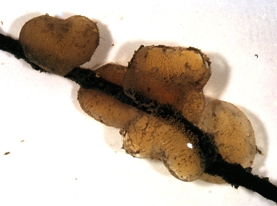
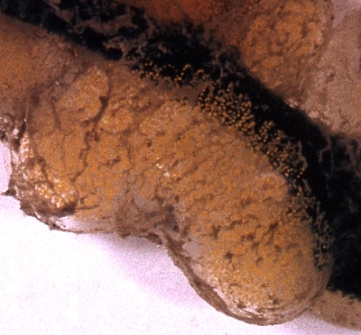
To accompany my other message on Haminoea zelandiae [#18126] here are some photos of its egg mass. All I can give as details is that each egg mass was 30 mm long and 10 mm in diameter. The egg masses are a very characteristic shape, consisting of a sausage mass of clear jelly in which is embedded a spiral string of bright yellow eggs.
Bill Rudman
Rudman, W.B., 2006 (Oct 27) Egg mass of Haminoea zelandiae. [Message in] Sea Slug Forum. Australian Museum, Sydney. Available from http://www.seaslugforum.net/find/18124Colour variability in Haminoea zelandiae
October 27, 2006
From: Bill Rudman
I came across these photos I took of Haminoea zelandiae many years ago. They show the range of colour variation from pale translucent brown to almost black. Unfortunately I wasn't very diligent in labelling slides in those days so I have no details on these photos other than that they would have been from somewhere in Auckland, New Zealand in 1969 or 1970 when I was studying their biology and comparative anatomy.
-
Rudman, W.B. (1971): On the opisthobranch genus Haminoea Turton & Kingston. Pacific Science, 25(4): 545-59, 12 figs.
-
Rudman, W.B. (1971) Structure and functioning of the gut in the bullomorpha (Opisthobranchia) Part 1. Herbivores. Journal of Natural History, 5(6): 647-675, 18 figs.
Bill Rudman
Haminoea zelandiae egg masses
December 8, 2003
From: Paul Furneaux
Dear Bill,
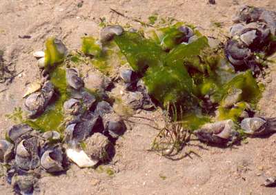
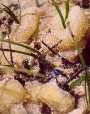
Although I've been busy with other species in Tauranga Harbour [North Island, New Zealand], Haminoea zelandiae has been busily reproducing throughout most of the year, but seemed to reach a peak during September when the proportion of large animals (50-60mm long) appeared to be greatest and the products of their reproduction, the jelly-like egg masses were incredibly numerous. In some situations the supply of Ulva was almost "cleaned up" by the large numbers of animals and almost any suitable surface was covered with their egg masses. By November the numbers of adult animals had diminished significantly leaving only their egg masses behind, although Haminoea is so abundant on these flats that it seems unlikely that there would ever be a period when there were no adult animals at all present. The photos show some of the massed feeding around a diminishing food supply, and some of the egg masses. These photos were taken on 7 October 2003.
I should have one more communication re. Bulla and one more re. Philinopsis before the Christmas break.
Regards,
Paul Furneaux.
P.Furneaux@xtra.co.nz
Dear Paul,
It's great to hear more about these animals. I agree that its hard to believe that animals so abundant can disappear at times, but that's exactly what happened the year I attempted to start a research project on Haminoea many years ago. They completely disappeared from the Auckland region for almost 18 months. I look forward to your messages on Bulla and Philinopsis.
Best wishes
Bill Rudman
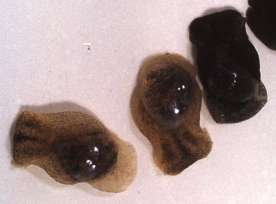
Haminoea zelandiae from Tauranga, New Zealand
June 6, 2003
From: Paul Furneaux
Hi Bill,
Thanks for your comments, the Philinopsis pix look great. I am including some Haminoea zelandiae photos with this message. They are extremely numerous where I have been working [Tauranga Harbour, east coast, Nth Island, New Zealand]. I have not done any counts of them but they would be from 10 to 30 per square metre in some situations, particularly in some patches of dense Zostera cover, and also in some areas of Ulva.
Both of these plants have been cause for concern in for quite different reasons, Zostera, because very large areas of it have disappeared over the past 40 - 50 years and thus a great deal of good habitat has been lost, and Ulva, because periodically it grows so extensively that it accumulates in quite thick layers or piles and becomes a problem in some localities. However both plants seem to be important to Haminoea zelandiae. I have one photo of an individual that is feeding on Ulva, " holding" it edge on, and they also appear to graze the algae that grow on the surface of the Zostera leaves. Their egg masses are also extremely common.
I have not yet found a live example of Bulla but their empty shells are reasonably common so perhaps a nocturnal excursion is called for.
Regards,
Paul Furneaux.
P.Furneaux@xtra.co.nz
Thanks Paul,
Photos of H. zelandiae certainly bring back memories. It is certainly one of the commonest sea slugs in New Zealand. It feeds on both the microalgal film growing on the leaves of Zostera and also on similar films or layers on soft sediments and hard surfaces. It also eats Ulva and 'filamentous' algae such as Enteromorpha. Actually it completely lacks self control when feeding and certainly in aquarium conditions it would eat Enteromorpha non-stop until it reached the stage where it would be defecating green pellets of undigested algae at one end while continuing to stuff in more food at the other end.
Your photos show the range in colour of this species from almost pure black to a light mottled brownish colour. The upper right photo of an animal ploughing through the sand, catching the fine sediment in the mucus sheet which covers its headshield, is a good example of how the headshield and its anterior mucus glands work together to allow the animal to live in these silty environments without clogging up the organs of the mantle cavity.
Best wishes,
Bill Rudman.
Shells of Haminoea zelandiae
June 6, 2003
From: Bill Rudman
Here is a scan of the shells of some Haminoea zelandiae I collected in 1968 in Auckland, New Zealand.
Herne Bay, Waitemata Hbr, Auckland, New Zealand. Pools in reef, 26 Sept. 1968. Shell length to 22 mm. AM C159944 [Cited Rudman 1971].
• Rudman, W.B. (1971) Structure and functioning of the gut in the bullomorpha (Opisthobranchia) Part 1. Herbivores. Journal of Natural History, 5(6): 647-675, 18 figs.
Bill Rudman
Rudman, W.B., 2003 (Jun 6) Shells of Haminoea zelandiae. [Message in] Sea Slug Forum. Australian Museum, Sydney. Available from http://www.seaslugforum.net/find/10168Gizzard plates of Haminoea zelandiae
December 23, 1999
From: Bill Rudman
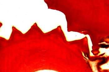
I don't have a photo of a typical Haminoea gizzard plate to compare with Clay & Patty Jo's photos of the gizzard plate of Atys naucum but here is a section through the gizzard of the common New Zealand species Haminoea zelandiae. In this section 2 of the 3 plates are visible, cut longitudinally. The lower plate is cut through about the midline and the upper more obliquely. The section clearly shows how the transverse ridges mesh together to form a very efficient crushing organ, grinding algal material to release the cell contents. The 8 or 9 ridges in species of Haminoea are quite different from the multitude of fine ridges in Atys naucum.
Bill Rudman.