Charcotia granulosa from Antarctica
February 22, 2007
From: David Cothran


Hi Bill,
I am a long term admirer of the Sea Slug Forum - it has been a fantastically useful resource for my work a marine naturalist for Lindblad Expeditions.
I have been diving in the Antarctic for the past 6 Austral summers and thought you might enjoy seeing a couple of the nudibranchs I have photographed; one of them has been puzzling me.
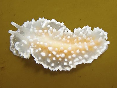
Locality [for 1st image]: Cuverville Island, Antarctic Peninsula, Southern Ocean, 12 meters. Length: 25 mm. 8 February 2007. Rock wall. Photos: David Cothran
The first image is, I believe, Charcotia granulosa, a moderately common species around the Antarctic Peninsula, relatively small and inconspicuous except for its white color.
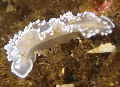
The second image is one I took at first to be another C. granulosa. On close examination though, I noticed a number of odd characteristics, including the form of the rhinophores, the web of orange veins and the reddish dots associated with them. I have photographed a number of C. granulosa specimens and never seen these characters before.
What do you think?
Best regards,
David Cothran
david@wandering-eye.com
Cothran, D., 2007 (Feb 22) Charcotia granulosa from Antarctica. [Message in] Sea Slug Forum. Australian Museum, Sydney. Available from http://www.seaslugforum.net/find/19508
Dear David,
Thanks very much for these interesting additions to the Forum. I think it's worth mentioning at this stage that our knowledge of the Antarctic opisthobranchs is based on a little preserved material and a little more recently collected material, some of which includes information on the living animals. Our biggest problem then is matching the original material, which was usually described from preserved, often distorted material, and lacks information on the shape and colour of the live animals, with more recently collected material, or photos like yours.
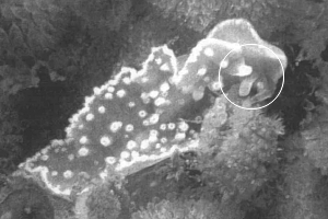
Your upper animal is almost certainly Charcotia granulosa, which was described from a single preserved specimen (Vayssiere, 1906). More recently, a few living animals were found and described (Wagele et al, 1995) and they look very similar to your photo. However one major difference is that they describe the rhinophores as 'petal-like, laterally compressed; tips digitiform', which seems rather different from the long smooth cylindrical rhinophores in your photo. Looking at their photo [see black & white copy alongside] it seems that the laterally compressed basal part of the rhinophores in their specimen may represent a partially contracted state. This example certainly illustrates the fairly primitive state of our knowledge. Any photos and information you can send will be of great value.
I think I will deal with your second photo in a separate message [#19509]. Although it has similarities to Charcotia, in that genus there are a few branches of the digestive gland running up to the the mantle edge but never into the oral veil and foot. However you can see in your photo that the brown branches of the digestive gland extend into the oral veil and down along the edge of the foot. I suspect it is Pseudotritonia quadrangularis but I will discuss that separately
One thing it would be nice to get a photo of is the right side of the body of either, or both of these species. Species of this family have a glandular band running along the right side of the body from the genital openings back toward the tail. I have no idea what it looks like in the living animal but I suspect it will be an opaque white line.
Best wishes,
Bill Rudman
Related messages
-
Re: Charcotia granulosa from the Antarctic Peninsula
From: Kevin Lee, March 19, 2010 -
Charcotia granulosa from the Antarctic Peninsula
From: Kevin Lee, March 18, 2010 -
Charcotia granulosa from Antarctica
From: Erling Svensen, January 27, 2010
